Posts

Showing posts with the label Oral Medicine & Oral Pathology

Oral Ulcers: Clinical features, Causes & Treatment

Image
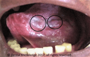
Traumatic ulcers in healing stage caused by sharp teeth An ulcer is a tissue defect which has penetrated the epithelial-connective tissue border, with its base at a deep level in the submucosa, or even within muscle or periosteum. An ulcer is a deeper breach of the epithelium than an erosion or an excoriation, and involves damage to both epithelium and lamina propria.

Introduction to Third Molar Surgery: Part-2 Indications and Contraindication

Image
Mandibular Third Molars   According to George Dimitroulis, there are common and uncommon reasons for the removal of the mandibular third molar.    

Oral Lichen Planus

Image
Oral Lichen Planus on left mucosa [1] Question: What is oral lichen planus? Answer: It is a chronic inflammatory condition that affects the skin, nails, hair, and mucous membranes, characterised by purplish, itchy, flat  eruptions. Question: How common is the condition? Answer: It is a common condition in India. Its cases are reported more than 10 lakh per year in India.   Question: How much time does it need for recovery? Answer: It can last several years or remains lifelong. Question: Is the condition treatable?   Answer: Treatments can help manage conditions. There is no known cure present.     Question: Does diagnosis require lab tests or imaging?   Answer: Its diagnosis rarely requires lab tests or imaging.         Condition Highlights   It commonly occurs for ages 35-50.   It is more common in females.   Family history may increase likelihood to occur.    

Cysts of the Jaws and Neck: Classification

  Cysts of the Jaws and Neck   Cysts can be classified in three types. Odontogenic Cysts   Periapical (Radicular) Cyst    Lateral Periodontal Cyst    Gingival Cyst of the Newborn    Dentigerous Cyst    Eruption Cyst    Glandular Odontogenic Cyst    Odontogenic  Keratocyst     Calcifying Odontogenic Cyst    Nonodontogenic Cysts   Globulomaxillary  Lesion    Nasolabial Cyst    Median Mandibular Cyst    Nasopalatine Canal Cyst    Pseudocysts   Aneurysmal Bone Cyst    Traumatic (Simple) Bone Cyst    Static Bone Cyst (Stafne’s Bone Defect)    Focal Osteoporotic Bone Marrow   Defect    Soft Tissue Cysts of the Neck    Branchial Cyst/ Cervical Lymphoepithelial   Cyst   

Dentigerous/Follicular Cysts

Image
Dentigerous (Follicular) Cysts are the second most commonly occurring odontogenic cysts after periapical cyst and the most common developmental cysts of the jaws. By definition, a dentigerous cyst is attached to the tooth cervix (enamel-cementum junction) and encloses the crown of the unerupted tooth. Photograph1: Dentigerous cyst surrounding the crown of right mandibular third molar and going upward in ascending ramus. [1] Etiology and Pathogenesis of Dentigerous Cyst   A dentigerous cyst originates  from the  enamel organ remnant or reduced enamel epithelium. The expansion of the dentigerous cyst is related to epithelial proliferation, release of bone-resorbing factors, and an increase in cyst fluid osmolality.   Clinical Features of Dentigerous Cyst   Dentigerous cysts are most commonly seen associated with third molars and maxillary Canines. The peak incidence of dentigerous cysts occurs between twenty to 40 ...

Odontogenic Keratocyst

Image
Odontogenic Keratocyst     Odontogenic keratocyst (OKC) is a parakeratin lined cyst like lesion within bone. OKCs mostly occur in the 2 nd and 3 rd decades of life; although can occur over a wide age range with male predilection. Odontogenic keratocyst comprises of 4 to 12 percent of all odontogenic cysts. Ninety percent of odontogenic keratocysts are solitary. Multiple cysts are found in Nevoid Basal Cell Carcinoma Syndrome / Gorlin Syndrome[1].   The most commonly involved body part in mandible, 65-85%; mostly posterior mandible. Normally, they are associated with third molars but may be present without associating with third molars; rarely occur in soft tissues. They are considered to arise from dental lamina.   Clinical Features   Odontogenic keratocysts are often asymptomatic. They are incidentally discovered on radiographs. They can cause symptomatic swelling. They can cause parasthesia of lip and teeth. Symptoms of pain and drainage are found if s...